What’s Hot

diabetes mellitus
Diabetes
A condition in which excessive amounts of some substances are excreted from the body. The term may refer to either of two unrelated diseases, diabetes mellitus and diabetes insipidus. The word diabetes derives from the Greek for siphon, a reference to the copious urine excretion that characterizes this affliction. In common usage, the term diabetes is synonymous with diabetes mellitus.
Diabetes Mellitus
Diabetes mellitus is a disease of abnormal carbohydrate metabolism in which glucose cannot enter the body's cells and be utilized, and therefore remains in the blood in high concentrations. Mellitus is the Latin word meaning honey, and is applicable because the urine of patients with diabetes mellitus has a sweet taste, distinguishing this disease from diabetes insipidus, in which large volumes of dilute, almost colorless and tasteless urine are passed. In diabetes mellitus the excess sugar in the blood (hyperglycemia) leads to the excretion of sugar in the urine (glycosuria), a cardinal diagnostic symptom. Glycosuria in turn causes the excretion of large amounts of urine (polyuria), which results in dehydration and intense thirst (polydipsia). Although blood glucose is high, it cannot enter the appetite-regulating cells of the hypothalamus; hunger is therefore great, and the diabetic person tends to eat constantly (polyphagia). But because glucose cannot enter and nourish the cells, body tissues are subjected to the equivalent of starvation; rapid weight loss occurs, part of which is due to the excretion of water in urine. See also: Carbohydrate metabolism
diabetes mellitus
Varieties
Diabetes mellitus appears in two varieties, each with its own cause: diabetes mellitus type I (formerly known as juvenile onset diabetes), caused by deficiency of the pancreatic hormone insulin (whose chief function is to promote the entry of glucose into cells); and diabetes mellitus type II (formerly known as maturity onset diabetes), in which insulin is available but cannot be properly utilized.
Diabetes mellitus type 1
diabetes mellitus type 1
Of the two forms of diabetes, type I is initially the more serious and less common, afflicting approximately 1 in every 600 children. The incidence varies among countries. The highest incidence is seen in Finland and Sweden, and some of the lowest in Korea and Mexico. The United States and Canada are included among the highest. Although it usually appears before the age of 20, type I diabetes can strike at any age. It is caused by a significant shortage or complete lack of insulin secretion. The endocrine cells normally responsible for insulin production are found in the pancreas, but have nothing to do with the ordinary digestive functions of that organ. They occur as islands of endocrine tissue, called islets of Langerhans, scattered throughout the substance of the pancreas. The islets consist of at least three cell types, each of which secretes a different hormone important in regulating carbohydrate metabolism. The alpha cells produce glucagon, which elevates blood glucose; the beta cells produce insulin; and the delta cells produce somatostatin, a hormone that appears to participate in controlling the activity of the alpha and beta cells.
In type I diabetes the beta cells are destroyed, possibly by the attack of the body's own immune system or by virus infection; victims of type I require daily insulin injections to survive. Insulin must be injected rather than taken orally because it is inactivated by the enzymes of the digestive system. Without insulin therapy, a patient with type I diabetes will inevitably deteriorate into a condition of ketosis (presence in the blood of ketone bodies, the metabolic intermediates of fat metabolism) and acidosis (dangerously acidic blood pH). Ketoacidosis, as the combined condition is called, leads rapidly to coma and death in the untreated diabetic.
When Frederick G. Banting and Charles H. Best succeeded in purifying insulin from animal pancreatic tissue in 1921, type I diabetics were given their first chance at living a normal life-span. Today type I diabetics are routinely treated by injections of insulin. Human insulin became increasingly available in the 1980s through the use of recombinant deoxyribonucleic acid (DNA) technology. In this technique the gene specifying human insulin is spliced into the chromosome of a bacterium, and the billions of progeny bacteria then produce insulin as though it were one of their own bacterial proteins, thus serving as minifactories for the hormone. The bacterium used to produce insulin is Escherichia coli. Another DNA technique for producing insulin uses yeast instead of E. coli. Up to the early 1980s, insulin was extracted from cattle and pigs. This “foreign” insulin is still used, but has some unwanted side effects not seen or rarely seen with human insulin, therefore making human insulin the treatment of choice. One of the most important characteristics of insulin produced by genetic engineering, other than its being identical to human pancreatic insulin, is its limitless availability. See also: Genetic engineering; Insulin; Pancreas
Diabetes mellitus type 2
diabetes mellitus type 2
Type II is the more common form of diabetes mellitus, accounting for 8 out of every 10 cases. It usually appears after the age of 30, although an uncommon variant called type II diabetes of the young occasionally occurs in obese children and teenagers. The initial symptoms of type II diabetes are much less noticeable than those of type I, and the characteristic triad of polyuria, polydipsia, and polyphagia may even be entirely absent. For this reason, type II diabetes can exist undetected for dangerously long periods. Victims are not prone to the ketoacidosis and coma that threaten type I patients, and their disease can usually be managed without insulin injections. Thus two other names for type II diabetes are nonketosis-prone diabetes and noninsulin-dependent diabetes.
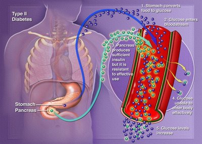
Whereas type I diabetes is a disease of insulin shortage, victims of type II diabetes usually have insulin in their bloodstream. In fact, insulin levels in type II diabetics are sometimes even higher than those in nondiabetic individuals. However, since the cells of a type II patient do not respond to insulin by taking in and utilizing blood glucose as normal cells would, the type II diabetic may have hyperglycemia in spite of high insulin levels. The probable reason for this defect is that the cells have specific receptor molecules on their surface membranes whose job is to recognize insulin and trigger the biochemical steps leading to glucose uptake and utilization. Interestingly, the number of insulin receptors on cells can be changed by gaining or losing body weight; the more body fat a person carries, the fewer insulin receptors there will be on the cells. This may explain why type II diabetes is mainly a disease of the obese, and why weight reduction is such effective therapy. High glucose levels also decrease the number of insulin receptors; normalizing glucose levels is thus of utmost importance.
The most important treatment for most cases of type II diabetes consists of exercise and weight reduction, which will return glucose metabolism to normal in 9 out of 10 cases. Because the cells of type II diabetics lack responsiveness to their own insulin, insulin injection therapy is used for some of these cases.
Approximately 10% of all type II patients must use insulin. This percentage appears to more than double in Black, Latino, Native American, or other minority patients living in the United States. In addition, a family of drugs known as oral hypoglycemic agents is often used in treating the disease. These drugs, including tolbutamide, chlorpropamide, acetohexamide, and tolazamide, lower blood glucose by stimulating the pancreatic cells to release insulin, and by augmenting insulin's effect on the cells of the body. All of these drugs are known as first-generation sulfonylureas. However, there are other drugs with the same basic molecule but with chemical changes in their structure; these are called second-generation sulfonylureas. This group of drugs is safe if properly used and is effective in many cases if used in conjunction with proper diet and exercise programs.
Amylin
Amylin is a 37-amino-acid peptide hormone that occurs naturally in humans, and has been shown to be in excess in individuals with non-insulin-dependent diabetes mellitus. Amylin has been shown to be deposited in the islets of Langerhans, and may decrease insulin release and insulin production in the pancreas, thus causing a deterioration in glucose control in non-insulin-dependent diabetes.
In insulin-dependent diabetes, amylin appears to slowly decrease with the progression of the disease, and when none is found, individuals may be more prone to have hypoglycemia unawareness (low blood glucose levels without symptoms).
Complications
Both type I and type II diabetes tend to produce a number of debilitating and life-threatening complications despite the use of insulin or oral hypoglycemic agents. It is primarily these complications, not the risk of ketoacidosis, that account for the heavy burden this disease inflicts on society, and make diabetes the third leading cause of death in the United States.
Much evidence indicates that most of the complications of diabetes are caused by the elevated blood glucose levels common in this disease. Even diabetics who take regular insulin injections exhibit wide fluctuations in their blood glucose levels, in marked contrast to the tightly controlled blood glucose levels observed in nondiabetics. According to the prevalent hypothesis, the more diabetics succeed in controlling their blood glucose to near-normal levels, the less risk there will be of developing any complications of the disease.
Thus, careful attention to diet, exercise, regular medication, and monitoring of blood glucose is vitally important for any diabetic.
The most common complications of diabetes are the following.
Eye damage
Diabetes is the leading cause of nontraumatic blindness in young adults. The blood vessels of the diabetic eye
are prone to microaneurysms (small outpouching of the vessel wall), excessive vascular permeability
(leakage from the vessel), growth of newer, more fragile vessels, and ultimately hemorrhage (bleeding).
These events can lead to scarring, fibrosis, and subsequent loss of vision.
Kidney damage
In long-standing diabetes, the smallest blood vessels of nephrons (the functional filtration units of the
kidney) frequently suffer the same type of damage as the blood vessels of the retina; this condition is called
diabetic nephropathy. Severe diabetic nephropathy commonly leads to kidney failure, and such patients
require dialysis in order to survive. The prognosis for individuals with diabetes on dialysis is poor, with only
30% surviving beyond 5 years.
Premature atherosclerosis
Atherosclerosis (often called arteriosclerosis or hardening of the arteries) is a condition in which arteries
become progressively occluded by accumulations of cholesterol-containing plaque. Although atherosclerosis is
common in older nondiabetics, diabetics tend to develop it earlier and in a more severe form. The most
common consequence of atherosclerosis is coronary artery disease and stroke, which result when the blood
supply to the heart or the brain is obstructed by a plaque within an artery. Coronary artery disease is the
leading cause of death in long-term diabetics, accounting for 75% of fatalities. Also, because of
atherosclerosis and restricted blood flow, diabetics are often subject to gangrene, especially of the
extremities. See also: Arteriosclerosis
Nervous system abnormalities
Patients with diabetes may experience a variety of nervous system disturbances, collectively referred to as
diabetic neuropathy. Symptoms may include muscle weakness, pain in the extremities, local paralysis,
urinary incontinence, sexual impotence, and sensations of cold, heat, or tingling in various parts of the body.
There may also be episodes of diarrhea, frequently nocturnal, which is a characteristic of the “diabetic
diarrhea” problems involving the upper intestinal tract. Nervous system involvement of the esophagus and
stomach may cause nausea and even vomiting.
Causes
Three factors that are believed to be important in the causation of this disease are heredity, viral infections,
and immunological damage to the pancreas.
Heredity
Although diabetes is popularly considered a genetic disease, it is not inherited in the sense that one can
inherit curly hair. Heredity does operate in determining one's risk of diabetes, but only as a predisposing
factor, not as an absolute determinant. The two types of diabetes mellitus have entirely different patterns of
inheritance, and the presence of one type in a family does not influence one's risk of developing the other.
Genetic factors play a much more important role in type II diabetes than in type I diabetes. For example, the
identical twin of a person who develops type II diabetes after age 40 is almost 100% certain to develop the
disease, but someone whose identical twin has type I diabetes runs only a 52% risk of becoming a diabetic.
Parents with type I diabetes face only a 2% risk of having a child with the disease, whereas the risk to the
offspring of a type II diabetic is about 10%. Genetic predisposition is apparently stronger in the case of type
II diabetes of the young, with a 50% chance that the child of such a diabetic will also develop the disease.
The environment obviously plays a significant role in type II diabetes. See also: Human genetics

Virus infections
Virus infection is strongly implicated as one causative factor in the beta-cell destruction that characterizes
type I diabetes, but it is not involved in type II diabetes. Some of the viruses that have been implicated are
the agents of mumps and rubella (German measles), whose action seems to be delayed for months or years,
and coxsackievirus B, which is more likely to precipitate diabetes shortly after the person recovers from the
influenzalike infection. Apparently, several factors operate to determine whether or not diabetes will develop
in a person infected with one of these extremely common viruses. They include the organ affinity of the
particular virus strain, the person's susceptibility to viral invasion of the pancreatic tissue, and the individual's
capacity to regenerate islet tissue that has been damaged by a viral infection. See also: Animal virus
Immunological damage to pancreas
Autoimmune reactions, wherein the body's immune defense system attacks its own pancreatic tissue as
though it were a foreign substance, are also suggested as a cause for the beta-cell destruction of type I
diabetes. An autoimmune response may be conducted by T lymphocytes or by soluble antibodies in the blood
or by both. These reactions may arise spontaneously or may be secondary to a viral infection of the pancreas
that leaves the islet tissue modified in some way that marks it for attack. See also: Autoimmunity
Diet
Dietary management has been part of diabetic therapy since preinsulin days, when starvation diets were
used to prolong the life of type I diabetics. More recently, weight reduction for victims of type II diabetes and
complex carbohydrate liberalization for those of both types of diabetes mellitus have been the cornerstone of
therapy.
For obese type II diabetics, weight reduction is certainly appropriate, since it tends to increase the number of insulin receptors on the cells of the body, usually making it possible to normalize blood glucose without
resorting to the use of insulin injections or oral drugs.
Some other dietary measures besides reducing caloric intake are now believed beneficial for type II diabetics.
The ingestion of foods high in fiber content, for example, has been shown to reduce hyperglycemia in type II
diabetics.
For type I diabetics, the traditional diet is high in protein and quite restricted in carbohydrates, with only
about 45% of the total daily caloric intake being carbohydrate-derived. Since most type I diabetics are thin,
weight reduction is not part of their program. Presently, the recommended carbohydrate intake has been
liberalized to 55–60% of the diet, plus 15–20% protein and 20–30% fat. There has been a trend toward
relaxing the carbohydrate restriction for type I diabetics as evidence accumulates that diets containing even
as much as 80% carbohydrates can be compatible with excellent control of diabetes, as long as the
carbohydrates are complex (that is, whole grains, legumes, potatoes, bread) rather than refined (that is,
sugars, candy, soft drinks, desserts, and so on). Artificially sweetened food can be very beneficial for weight
control; however, it is not a necessary component of treatment.
Prospects
The ideal solution is to be able to mimic insulin secretion, with intensive insulin therapy used with either
multiple insulin injections or insulin infusion pump therapy. The insulin pump continuously delivers a small
amount of insulin, and the individual can increase the amount before meals based on his or her glucose level.
Still, this setup is not ideal: in a nondiabetic, insulin is delivered directly into the portal blood system, while
therapy delivers insulin under the skin, where it can be absorbed into the bloodstream. Hooking a glucosemonitoring
device to the insulin pump would allow the system to close, permitting the individual to have an
artificial pancreas implanted (several models are available).
Healthy pancreatic tissue can be transplanted from a donor into a type I diabetic, but there are many
problems related to this method, such as the typing of the pancreatic tissue from the donor, the possibility of
rejection, and the possibility of infections.
diabetes mellitus
Diabetes Insipidus
Diabetes insipidus is caused by a deficiency of, or resistance to the action of, vasopressin, the antidiuretic
hormone produced by the posterior lobe of the pituitary gland. If the pituitary fails to produce vasopressin,
the condition is called central diabetes insipidus. If the kidneys do not respond to the vasopressin and fail to
concentrate urine, the condition is labeled nephrogenic diabetes insipidus. Symptoms include increased thirst
and frequent urination. Permanent damage to the kidneys can result if the condition is not treated.
A low level of vasopressin can be corrected with desmopressin acetate, a synthetic analog of 8-argininevasopressin,
which can be administered in the form of a nasal spray. Desmopressin acetate is ineffective
in the treatment of nephrogenic diabetes insipidus. See also: Neurohypophysis hormone
diabetes mellitus - diabetes mellitus type 2 - diabetes mellitus type 1

diabetes mellitus

diabetes mellitus
Diabetes
A condition in which excessive amounts of some substances are excreted from the body. The term may refer to either of two unrelated diseases, diabetes mellitus and diabetes insipidus. The word diabetes derives from the Greek for siphon, a reference to the copious urine excretion that characterizes this affliction. In common usage, the term diabetes is synonymous with diabetes mellitus.
Diabetes Mellitus
Diabetes mellitus is a disease of abnormal carbohydrate metabolism in which glucose cannot enter the body's cells and be utilized, and therefore remains in the blood in high concentrations. Mellitus is the Latin word meaning honey, and is applicable because the urine of patients with diabetes mellitus has a sweet taste, distinguishing this disease from diabetes insipidus, in which large volumes of dilute, almost colorless and tasteless urine are passed. In diabetes mellitus the excess sugar in the blood (hyperglycemia) leads to the excretion of sugar in the urine (glycosuria), a cardinal diagnostic symptom. Glycosuria in turn causes the excretion of large amounts of urine (polyuria), which results in dehydration and intense thirst (polydipsia). Although blood glucose is high, it cannot enter the appetite-regulating cells of the hypothalamus; hunger is therefore great, and the diabetic person tends to eat constantly (polyphagia). But because glucose cannot enter and nourish the cells, body tissues are subjected to the equivalent of starvation; rapid weight loss occurs, part of which is due to the excretion of water in urine. See also: Carbohydrate metabolism
diabetes mellitus
Varieties
Diabetes mellitus appears in two varieties, each with its own cause: diabetes mellitus type I (formerly known as juvenile onset diabetes), caused by deficiency of the pancreatic hormone insulin (whose chief function is to promote the entry of glucose into cells); and diabetes mellitus type II (formerly known as maturity onset diabetes), in which insulin is available but cannot be properly utilized.
Diabetes mellitus type 1
diabetes mellitus type 1
Of the two forms of diabetes, type I is initially the more serious and less common, afflicting approximately 1 in every 600 children. The incidence varies among countries. The highest incidence is seen in Finland and Sweden, and some of the lowest in Korea and Mexico. The United States and Canada are included among the highest. Although it usually appears before the age of 20, type I diabetes can strike at any age. It is caused by a significant shortage or complete lack of insulin secretion. The endocrine cells normally responsible for insulin production are found in the pancreas, but have nothing to do with the ordinary digestive functions of that organ. They occur as islands of endocrine tissue, called islets of Langerhans, scattered throughout the substance of the pancreas. The islets consist of at least three cell types, each of which secretes a different hormone important in regulating carbohydrate metabolism. The alpha cells produce glucagon, which elevates blood glucose; the beta cells produce insulin; and the delta cells produce somatostatin, a hormone that appears to participate in controlling the activity of the alpha and beta cells.
In type I diabetes the beta cells are destroyed, possibly by the attack of the body's own immune system or by virus infection; victims of type I require daily insulin injections to survive. Insulin must be injected rather than taken orally because it is inactivated by the enzymes of the digestive system. Without insulin therapy, a patient with type I diabetes will inevitably deteriorate into a condition of ketosis (presence in the blood of ketone bodies, the metabolic intermediates of fat metabolism) and acidosis (dangerously acidic blood pH). Ketoacidosis, as the combined condition is called, leads rapidly to coma and death in the untreated diabetic.
When Frederick G. Banting and Charles H. Best succeeded in purifying insulin from animal pancreatic tissue in 1921, type I diabetics were given their first chance at living a normal life-span. Today type I diabetics are routinely treated by injections of insulin. Human insulin became increasingly available in the 1980s through the use of recombinant deoxyribonucleic acid (DNA) technology. In this technique the gene specifying human insulin is spliced into the chromosome of a bacterium, and the billions of progeny bacteria then produce insulin as though it were one of their own bacterial proteins, thus serving as minifactories for the hormone. The bacterium used to produce insulin is Escherichia coli. Another DNA technique for producing insulin uses yeast instead of E. coli. Up to the early 1980s, insulin was extracted from cattle and pigs. This “foreign” insulin is still used, but has some unwanted side effects not seen or rarely seen with human insulin, therefore making human insulin the treatment of choice. One of the most important characteristics of insulin produced by genetic engineering, other than its being identical to human pancreatic insulin, is its limitless availability. See also: Genetic engineering; Insulin; Pancreas
Diabetes mellitus type 2
diabetes mellitus type 2
Type II is the more common form of diabetes mellitus, accounting for 8 out of every 10 cases. It usually appears after the age of 30, although an uncommon variant called type II diabetes of the young occasionally occurs in obese children and teenagers. The initial symptoms of type II diabetes are much less noticeable than those of type I, and the characteristic triad of polyuria, polydipsia, and polyphagia may even be entirely absent. For this reason, type II diabetes can exist undetected for dangerously long periods. Victims are not prone to the ketoacidosis and coma that threaten type I patients, and their disease can usually be managed without insulin injections. Thus two other names for type II diabetes are nonketosis-prone diabetes and noninsulin-dependent diabetes.
Whereas type I diabetes is a disease of insulin shortage, victims of type II diabetes usually have insulin in their bloodstream. In fact, insulin levels in type II diabetics are sometimes even higher than those in nondiabetic individuals. However, since the cells of a type II patient do not respond to insulin by taking in and utilizing blood glucose as normal cells would, the type II diabetic may have hyperglycemia in spite of high insulin levels. The probable reason for this defect is that the cells have specific receptor molecules on their surface membranes whose job is to recognize insulin and trigger the biochemical steps leading to glucose uptake and utilization. Interestingly, the number of insulin receptors on cells can be changed by gaining or losing body weight; the more body fat a person carries, the fewer insulin receptors there will be on the cells. This may explain why type II diabetes is mainly a disease of the obese, and why weight reduction is such effective therapy. High glucose levels also decrease the number of insulin receptors; normalizing glucose levels is thus of utmost importance.
The most important treatment for most cases of type II diabetes consists of exercise and weight reduction, which will return glucose metabolism to normal in 9 out of 10 cases. Because the cells of type II diabetics lack responsiveness to their own insulin, insulin injection therapy is used for some of these cases.
Approximately 10% of all type II patients must use insulin. This percentage appears to more than double in Black, Latino, Native American, or other minority patients living in the United States. In addition, a family of drugs known as oral hypoglycemic agents is often used in treating the disease. These drugs, including tolbutamide, chlorpropamide, acetohexamide, and tolazamide, lower blood glucose by stimulating the pancreatic cells to release insulin, and by augmenting insulin's effect on the cells of the body. All of these drugs are known as first-generation sulfonylureas. However, there are other drugs with the same basic molecule but with chemical changes in their structure; these are called second-generation sulfonylureas. This group of drugs is safe if properly used and is effective in many cases if used in conjunction with proper diet and exercise programs.
Amylin
Amylin is a 37-amino-acid peptide hormone that occurs naturally in humans, and has been shown to be in excess in individuals with non-insulin-dependent diabetes mellitus. Amylin has been shown to be deposited in the islets of Langerhans, and may decrease insulin release and insulin production in the pancreas, thus causing a deterioration in glucose control in non-insulin-dependent diabetes.
In insulin-dependent diabetes, amylin appears to slowly decrease with the progression of the disease, and when none is found, individuals may be more prone to have hypoglycemia unawareness (low blood glucose levels without symptoms).
Complications
Both type I and type II diabetes tend to produce a number of debilitating and life-threatening complications despite the use of insulin or oral hypoglycemic agents. It is primarily these complications, not the risk of ketoacidosis, that account for the heavy burden this disease inflicts on society, and make diabetes the third leading cause of death in the United States.
Much evidence indicates that most of the complications of diabetes are caused by the elevated blood glucose levels common in this disease. Even diabetics who take regular insulin injections exhibit wide fluctuations in their blood glucose levels, in marked contrast to the tightly controlled blood glucose levels observed in nondiabetics. According to the prevalent hypothesis, the more diabetics succeed in controlling their blood glucose to near-normal levels, the less risk there will be of developing any complications of the disease.
Thus, careful attention to diet, exercise, regular medication, and monitoring of blood glucose is vitally important for any diabetic.
The most common complications of diabetes are the following.
Eye damage
Diabetes is the leading cause of nontraumatic blindness in young adults. The blood vessels of the diabetic eye
are prone to microaneurysms (small outpouching of the vessel wall), excessive vascular permeability
(leakage from the vessel), growth of newer, more fragile vessels, and ultimately hemorrhage (bleeding).
These events can lead to scarring, fibrosis, and subsequent loss of vision.
Kidney damage
In long-standing diabetes, the smallest blood vessels of nephrons (the functional filtration units of the
kidney) frequently suffer the same type of damage as the blood vessels of the retina; this condition is called
diabetic nephropathy. Severe diabetic nephropathy commonly leads to kidney failure, and such patients
require dialysis in order to survive. The prognosis for individuals with diabetes on dialysis is poor, with only
30% surviving beyond 5 years.
Premature atherosclerosis
Atherosclerosis (often called arteriosclerosis or hardening of the arteries) is a condition in which arteries
become progressively occluded by accumulations of cholesterol-containing plaque. Although atherosclerosis is
common in older nondiabetics, diabetics tend to develop it earlier and in a more severe form. The most
common consequence of atherosclerosis is coronary artery disease and stroke, which result when the blood
supply to the heart or the brain is obstructed by a plaque within an artery. Coronary artery disease is the
leading cause of death in long-term diabetics, accounting for 75% of fatalities. Also, because of
atherosclerosis and restricted blood flow, diabetics are often subject to gangrene, especially of the
extremities. See also: Arteriosclerosis
Nervous system abnormalities
Patients with diabetes may experience a variety of nervous system disturbances, collectively referred to as
diabetic neuropathy. Symptoms may include muscle weakness, pain in the extremities, local paralysis,
urinary incontinence, sexual impotence, and sensations of cold, heat, or tingling in various parts of the body.
There may also be episodes of diarrhea, frequently nocturnal, which is a characteristic of the “diabetic
diarrhea” problems involving the upper intestinal tract. Nervous system involvement of the esophagus and
stomach may cause nausea and even vomiting.
Causes
Three factors that are believed to be important in the causation of this disease are heredity, viral infections,
and immunological damage to the pancreas.
Heredity
Although diabetes is popularly considered a genetic disease, it is not inherited in the sense that one can
inherit curly hair. Heredity does operate in determining one's risk of diabetes, but only as a predisposing
factor, not as an absolute determinant. The two types of diabetes mellitus have entirely different patterns of
inheritance, and the presence of one type in a family does not influence one's risk of developing the other.
Genetic factors play a much more important role in type II diabetes than in type I diabetes. For example, the
identical twin of a person who develops type II diabetes after age 40 is almost 100% certain to develop the
disease, but someone whose identical twin has type I diabetes runs only a 52% risk of becoming a diabetic.
Parents with type I diabetes face only a 2% risk of having a child with the disease, whereas the risk to the
offspring of a type II diabetic is about 10%. Genetic predisposition is apparently stronger in the case of type
II diabetes of the young, with a 50% chance that the child of such a diabetic will also develop the disease.
The environment obviously plays a significant role in type II diabetes. See also: Human genetics

Virus infections
Virus infection is strongly implicated as one causative factor in the beta-cell destruction that characterizes
type I diabetes, but it is not involved in type II diabetes. Some of the viruses that have been implicated are
the agents of mumps and rubella (German measles), whose action seems to be delayed for months or years,
and coxsackievirus B, which is more likely to precipitate diabetes shortly after the person recovers from the
influenzalike infection. Apparently, several factors operate to determine whether or not diabetes will develop
in a person infected with one of these extremely common viruses. They include the organ affinity of the
particular virus strain, the person's susceptibility to viral invasion of the pancreatic tissue, and the individual's
capacity to regenerate islet tissue that has been damaged by a viral infection. See also: Animal virus
Immunological damage to pancreas
Autoimmune reactions, wherein the body's immune defense system attacks its own pancreatic tissue as
though it were a foreign substance, are also suggested as a cause for the beta-cell destruction of type I
diabetes. An autoimmune response may be conducted by T lymphocytes or by soluble antibodies in the blood
or by both. These reactions may arise spontaneously or may be secondary to a viral infection of the pancreas
that leaves the islet tissue modified in some way that marks it for attack. See also: Autoimmunity
Diet
Dietary management has been part of diabetic therapy since preinsulin days, when starvation diets were
used to prolong the life of type I diabetics. More recently, weight reduction for victims of type II diabetes and
complex carbohydrate liberalization for those of both types of diabetes mellitus have been the cornerstone of
therapy.
For obese type II diabetics, weight reduction is certainly appropriate, since it tends to increase the number of insulin receptors on the cells of the body, usually making it possible to normalize blood glucose without
resorting to the use of insulin injections or oral drugs.
Some other dietary measures besides reducing caloric intake are now believed beneficial for type II diabetics.
The ingestion of foods high in fiber content, for example, has been shown to reduce hyperglycemia in type II
diabetics.
For type I diabetics, the traditional diet is high in protein and quite restricted in carbohydrates, with only
about 45% of the total daily caloric intake being carbohydrate-derived. Since most type I diabetics are thin,
weight reduction is not part of their program. Presently, the recommended carbohydrate intake has been
liberalized to 55–60% of the diet, plus 15–20% protein and 20–30% fat. There has been a trend toward
relaxing the carbohydrate restriction for type I diabetics as evidence accumulates that diets containing even
as much as 80% carbohydrates can be compatible with excellent control of diabetes, as long as the
carbohydrates are complex (that is, whole grains, legumes, potatoes, bread) rather than refined (that is,
sugars, candy, soft drinks, desserts, and so on). Artificially sweetened food can be very beneficial for weight
control; however, it is not a necessary component of treatment.
Prospects
The ideal solution is to be able to mimic insulin secretion, with intensive insulin therapy used with either
multiple insulin injections or insulin infusion pump therapy. The insulin pump continuously delivers a small
amount of insulin, and the individual can increase the amount before meals based on his or her glucose level.
Still, this setup is not ideal: in a nondiabetic, insulin is delivered directly into the portal blood system, while
therapy delivers insulin under the skin, where it can be absorbed into the bloodstream. Hooking a glucosemonitoring
device to the insulin pump would allow the system to close, permitting the individual to have an
artificial pancreas implanted (several models are available).
Healthy pancreatic tissue can be transplanted from a donor into a type I diabetic, but there are many
problems related to this method, such as the typing of the pancreatic tissue from the donor, the possibility of
rejection, and the possibility of infections.
diabetes mellitus
Diabetes Insipidus
Diabetes insipidus is caused by a deficiency of, or resistance to the action of, vasopressin, the antidiuretic
hormone produced by the posterior lobe of the pituitary gland. If the pituitary fails to produce vasopressin,
the condition is called central diabetes insipidus. If the kidneys do not respond to the vasopressin and fail to
concentrate urine, the condition is labeled nephrogenic diabetes insipidus. Symptoms include increased thirst
and frequent urination. Permanent damage to the kidneys can result if the condition is not treated.
A low level of vasopressin can be corrected with desmopressin acetate, a synthetic analog of 8-argininevasopressin,
which can be administered in the form of a nasal spray. Desmopressin acetate is ineffective
in the treatment of nephrogenic diabetes insipidus. See also: Neurohypophysis hormone
diabetes mellitus - diabetes mellitus type 2 - diabetes mellitus type 1

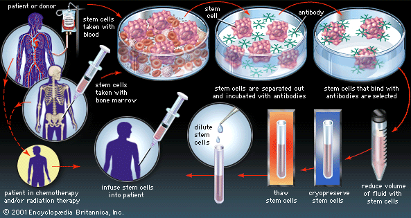
What is bone marrow transplantation - what is bone marrow transplant for donor
Bone marrow transplantation is a procedure in which bone marrow cells are taken out from a donor and given to a patient.
Depending on the donor or source of bone marrow, different types of bone marrow transplantation is done.
There are two major ways in which bone marrow cells can be removed from a person or donor. The first is by taking the bone marrow cells directly from the marrow using long needles (called bone marrow harvest needles) to suck out the marrow from the marrow spaces at the large hipbone. This is done under anaesthesia over about 2 hours. The second is a revolutionary method, which have been available only in the last 5-10 years. By giving certain medications, the bone marrow cells can be stimulated to circulate from the marrow into the blood. Using a special machine called an apheresis machine ( also called a cell separator), the cells can be removed from the blood. Blood is drawn out as in a blood donation, into the machine, which then separates out the marrow cells and return blood cells to the donor.
The donor is usually a relative, usually either a brother or sister. However, there are three other sources. They are unrelated match donor, cord blood and autologous.
Unrelated donors are donors that have volunteered to donate bone marrow just like blood donors.
Cord blood is obtained from the cord of newborn babies. It may be used for transplantation of a relative or be used for non-related recipients.
It is also possible for the patient to donate his own marrow cells to be stored first and then returned to him after the chemotherapy has been given as conditioning.
(See autologous BMT section)
What is bone marrow transplant
Figure 22b: Sources of haemopoeitic stem cells for transplantation
Sibling (Brother and sisters)   This is commonest form in the allogeneic setting.
Twin     An identical twin shares the same ‘marrow type’ as the patient, thus making it very safe.

Extended family  Sometimes a family member may share the same ‘marrow type’ as the patient and can donate to the patient.
Unrelated donors Volunteer donors who have the same ‘marrow type’ are able to donate their marrow for needy patients.
Autologous The patient’s own marrow can be used to do a transplant. (See chapter on autologous transplantation)
Thus the different types of transplants are:
Allogeneic bone marrow transplantation Allogeneic peripheral stem cell transplantation Autologous bone marrow transplantation Autologous peripheral stem cell transplantation Cord blood transplantation.
Who should receive a bone marrow transplant?
There are three important conditions limiting the use of bone marrow transplantation:
(i) Age - The older the patient, the more risky the transplant because of a higher incidence of complications. The upper age limit of transplants locally is currently 40 years of age. In some overseas centres, the upper age limit is 55 years of age.
(ii) Suitable disease - Bone marrow transplantation is not suitable for all blood diseases. The results are best for acute myeloid leukaemia if the transplant is done only when the patient has achieved remission. In chronic myeloid leukaemia the results are best when the transplant is performed relatively early i.e. when the patient is still in the chronic phase of the disease. In severe aplastic anaemia, new marrow is required to replace the patient's marrow that has completely failed.
(iii) Suitable donor - Allogeneic bone marrow transplants require a donor with the same tissue type as the patient. There is no age limit for donors. However the donor must be healthy and has not transmissible diseases.
How to find a suitable donor?
Our immune system which protects us from bacteria, viruses and other microorganisms which try to invade our bodies, has to have a way of identifying which cells are our own and which cells (micro-organisms) are ‘enemies’ that has to be destroyed. They are able to recognise our own cells because the cells have antigens (markers) on their surfaces. The antigens that we know are called HLA antigens. (HLA stands for human leucocyte antigens.) An analogy can be made. In war we can recognise each other by the uniforms or badges we wear. The enemy wears different uniforms and thus are easily recognised and attacked. We will not attack our own soldiers who are wearing the same uniforms.
These HLA antigens are unique in that they differ in us from one another. Family members tend to share same antigens. Brothers and sisters have a high chance of having the same antigens.
The process in which we check to see what antigens are carried on our cells is called tissue typing. Thus we are able to find out one’s tissue type using this process. This process is also called ‘HLA typing’.
What is bone marrow transplantation
How is tissue typing done?
A bone marrow test is not required. Only blood is needed because the white cells (mainly lymphocytes) that is in our blood is used. A blood sample is taken and using sophisticated tests the tissue type is determined.
In Malaysia, tissue typing is performed by the Institute of Medical Research, Kuala Lumpur and recently in University Hospital, Kuala Lumpur. Singapore is the nearest centre where this is also done. Only blood samples in special bottles need to be sent and the donors and patients do not need to go to the centres themselves.
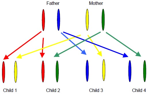
The chance of finding a suitable donor is best from a brother or a sister of the patient. There is a one in four chance of a brother or a sister having a complete match. The chances of the rest of the family (parents or children) having the same tissue type are much less (about one in 100). This is because genes inherited from
both parents of the patient determine the tissue or HLA type. The same tissue type is thus most likely to be found among the patient’s brothers and sisters (siblings).
Different ethnic groups have different HLA types just like hair color or facial features.
Thus if it is more likely to find the same tissue type in persons of the same ethnicity.
Figure 23a: Inheritance of HLA antigens
There are four possibilities for the type of HLA inherited from parents. They are inherited in pairs. We obtain one of the pairs from each parent. Thus the chances of having a sibling with the same tissue type is about 25%.
What is bone marrow transplantation
What are the problems associated with bone marrow transplants?
Sterility - the inability to have children is a permanent side effect of bone marrow transplantation. The ability to participate in sexual activity is not affected. In overseas centres, sperm banking is an option for male patients. Also eggs may also be stored for the female.
Skin changes and hair loss- the hair loss is only temporary and the hair will grow back after the transplant but sometimes is a bit thinner than previously. There are skin changes due to the chemotherapy given for the transplant but these are also temporary.
Mucositis - during the transplant, most patients develop a very sore mouth from the chemotherapy and for a while, cannot eat and will depend on nutrition given intravenously. Pain relieve can be achieved usually with medication.
Graft-versus-host-disease - this is a special complication of transplants due to the immune cells (lymphocytes) from the donor present in the donated marrow, which attack the recipient (patient). This is because they recognise the patient’s body cells as foreign like bacteria or micro-organisms. This occurs more often in HLA
mismatched transplants and unrelated donor transplants. It can also occur in fully matched transplants because there are still antigens that we yet cannot tissue type.
Also they may be other factors that change the donated cells resulting in them not recognising the shared HLA antigens as shared.
It can cause rashes, liver damage and damage to the gut. It occurs in about 40% of patients but can usually be treated. It is occasionally fatal.
Infections - During and after transplants, the patient remains at high risk of infections and will be on antibiotics and special medication for about one year. The immunity does not return to normal for a few years.
What does the patient undergo?
Because the procedure is very tedious and also difficult, the donor is counselled at length and allowed to make his or her own mind to go ahead with the transplantation.
The transplant patient is hospitalised for about one month but will require very close follow up at least three times a week for the subsequent two months. During the hospitalisation, a special device called a Hickman catheter is placed (using minor surgery) which serves as a way of giving fluids, chemotherapy, blood transfusion and nutrition directly into the bloodstream. This eliminates the need to use veins on
the arms to take blood samples and give intravenous medication.
The patient undergoes conditioning chemotherapy to prepare the patient to receive the new marrow. Conditioning chemotherapy has side effects including vomiting, diarrhoea and occasional bleeding from the bladder. The new marrow collected from the donor is then transfused into the patient. These cells know how to get to the bone marrow spaces through the bloodstream in a normal process called homing. The blood counts of the patient remain very low for about 2 to 3 weeks before the new marrow grows. The patient will be required to take medications for at least a year.
What does the donor undergo?
The donor is counselled (given advice) about the procedure and how it is useful for the patient. As the procedure is completely voluntary, the donor’s wishes are highly respected.
Depending on the procedure to be used whether conventional bone marrow harvest or peripheral stem cell collection, the donor is selected.
In the conventional bone marrow harvest programmes, the donor undergoes a medical examination to determine fitness for surgery. A unit of blood may be taken a week prior to the harvest and given back to the donor at the time of harvest so as to minimise the need for additional blood transfusion. The harvest is done under general anaesthesia and marrow is aspirated using special needles from the back of the pelvis (hip bone). There is no permanent side effect and the main complication is some discomfort felt at the back for several days after the procedure. The donor is usually hospitalised for three days only.
what is bone marrow transplant for - what is bone marrow transplant for donor - what is bone marrow transplant like

What is bone marrow transplant

What is bone marrow transplantation - what is bone marrow transplant for donor
Bone marrow transplantation is a procedure in which bone marrow cells are taken out from a donor and given to a patient.
Depending on the donor or source of bone marrow, different types of bone marrow transplantation is done.
There are two major ways in which bone marrow cells can be removed from a person or donor. The first is by taking the bone marrow cells directly from the marrow using long needles (called bone marrow harvest needles) to suck out the marrow from the marrow spaces at the large hipbone. This is done under anaesthesia over about 2 hours. The second is a revolutionary method, which have been available only in the last 5-10 years. By giving certain medications, the bone marrow cells can be stimulated to circulate from the marrow into the blood. Using a special machine called an apheresis machine ( also called a cell separator), the cells can be removed from the blood. Blood is drawn out as in a blood donation, into the machine, which then separates out the marrow cells and return blood cells to the donor.
The donor is usually a relative, usually either a brother or sister. However, there are three other sources. They are unrelated match donor, cord blood and autologous.
Unrelated donors are donors that have volunteered to donate bone marrow just like blood donors.
Cord blood is obtained from the cord of newborn babies. It may be used for transplantation of a relative or be used for non-related recipients.
It is also possible for the patient to donate his own marrow cells to be stored first and then returned to him after the chemotherapy has been given as conditioning.
(See autologous BMT section)
What is bone marrow transplant
Figure 22b: Sources of haemopoeitic stem cells for transplantation
Sibling (Brother and sisters)   This is commonest form in the allogeneic setting.
Twin     An identical twin shares the same ‘marrow type’ as the patient, thus making it very safe.

Extended family  Sometimes a family member may share the same ‘marrow type’ as the patient and can donate to the patient.
Unrelated donors Volunteer donors who have the same ‘marrow type’ are able to donate their marrow for needy patients.
Autologous The patient’s own marrow can be used to do a transplant. (See chapter on autologous transplantation)
Thus the different types of transplants are:
Allogeneic bone marrow transplantation Allogeneic peripheral stem cell transplantation Autologous bone marrow transplantation Autologous peripheral stem cell transplantation Cord blood transplantation.
Who should receive a bone marrow transplant?
There are three important conditions limiting the use of bone marrow transplantation:
(i) Age - The older the patient, the more risky the transplant because of a higher incidence of complications. The upper age limit of transplants locally is currently 40 years of age. In some overseas centres, the upper age limit is 55 years of age.
(ii) Suitable disease - Bone marrow transplantation is not suitable for all blood diseases. The results are best for acute myeloid leukaemia if the transplant is done only when the patient has achieved remission. In chronic myeloid leukaemia the results are best when the transplant is performed relatively early i.e. when the patient is still in the chronic phase of the disease. In severe aplastic anaemia, new marrow is required to replace the patient's marrow that has completely failed.
(iii) Suitable donor - Allogeneic bone marrow transplants require a donor with the same tissue type as the patient. There is no age limit for donors. However the donor must be healthy and has not transmissible diseases.
How to find a suitable donor?
Our immune system which protects us from bacteria, viruses and other microorganisms which try to invade our bodies, has to have a way of identifying which cells are our own and which cells (micro-organisms) are ‘enemies’ that has to be destroyed. They are able to recognise our own cells because the cells have antigens (markers) on their surfaces. The antigens that we know are called HLA antigens. (HLA stands for human leucocyte antigens.) An analogy can be made. In war we can recognise each other by the uniforms or badges we wear. The enemy wears different uniforms and thus are easily recognised and attacked. We will not attack our own soldiers who are wearing the same uniforms.
These HLA antigens are unique in that they differ in us from one another. Family members tend to share same antigens. Brothers and sisters have a high chance of having the same antigens.
The process in which we check to see what antigens are carried on our cells is called tissue typing. Thus we are able to find out one’s tissue type using this process. This process is also called ‘HLA typing’.
What is bone marrow transplantation
How is tissue typing done?
A bone marrow test is not required. Only blood is needed because the white cells (mainly lymphocytes) that is in our blood is used. A blood sample is taken and using sophisticated tests the tissue type is determined.
In Malaysia, tissue typing is performed by the Institute of Medical Research, Kuala Lumpur and recently in University Hospital, Kuala Lumpur. Singapore is the nearest centre where this is also done. Only blood samples in special bottles need to be sent and the donors and patients do not need to go to the centres themselves.
The chance of finding a suitable donor is best from a brother or a sister of the patient. There is a one in four chance of a brother or a sister having a complete match. The chances of the rest of the family (parents or children) having the same tissue type are much less (about one in 100). This is because genes inherited from
both parents of the patient determine the tissue or HLA type. The same tissue type is thus most likely to be found among the patient’s brothers and sisters (siblings).
Different ethnic groups have different HLA types just like hair color or facial features.
Thus if it is more likely to find the same tissue type in persons of the same ethnicity.
Figure 23a: Inheritance of HLA antigens
There are four possibilities for the type of HLA inherited from parents. They are inherited in pairs. We obtain one of the pairs from each parent. Thus the chances of having a sibling with the same tissue type is about 25%.
What is bone marrow transplantation
What are the problems associated with bone marrow transplants?
Sterility - the inability to have children is a permanent side effect of bone marrow transplantation. The ability to participate in sexual activity is not affected. In overseas centres, sperm banking is an option for male patients. Also eggs may also be stored for the female.
Skin changes and hair loss- the hair loss is only temporary and the hair will grow back after the transplant but sometimes is a bit thinner than previously. There are skin changes due to the chemotherapy given for the transplant but these are also temporary.
Mucositis - during the transplant, most patients develop a very sore mouth from the chemotherapy and for a while, cannot eat and will depend on nutrition given intravenously. Pain relieve can be achieved usually with medication.
Graft-versus-host-disease - this is a special complication of transplants due to the immune cells (lymphocytes) from the donor present in the donated marrow, which attack the recipient (patient). This is because they recognise the patient’s body cells as foreign like bacteria or micro-organisms. This occurs more often in HLA
mismatched transplants and unrelated donor transplants. It can also occur in fully matched transplants because there are still antigens that we yet cannot tissue type.
Also they may be other factors that change the donated cells resulting in them not recognising the shared HLA antigens as shared.
It can cause rashes, liver damage and damage to the gut. It occurs in about 40% of patients but can usually be treated. It is occasionally fatal.
Infections - During and after transplants, the patient remains at high risk of infections and will be on antibiotics and special medication for about one year. The immunity does not return to normal for a few years.
What does the patient undergo?
Because the procedure is very tedious and also difficult, the donor is counselled at length and allowed to make his or her own mind to go ahead with the transplantation.
The transplant patient is hospitalised for about one month but will require very close follow up at least three times a week for the subsequent two months. During the hospitalisation, a special device called a Hickman catheter is placed (using minor surgery) which serves as a way of giving fluids, chemotherapy, blood transfusion and nutrition directly into the bloodstream. This eliminates the need to use veins on
the arms to take blood samples and give intravenous medication.
The patient undergoes conditioning chemotherapy to prepare the patient to receive the new marrow. Conditioning chemotherapy has side effects including vomiting, diarrhoea and occasional bleeding from the bladder. The new marrow collected from the donor is then transfused into the patient. These cells know how to get to the bone marrow spaces through the bloodstream in a normal process called homing. The blood counts of the patient remain very low for about 2 to 3 weeks before the new marrow grows. The patient will be required to take medications for at least a year.
What does the donor undergo?
The donor is counselled (given advice) about the procedure and how it is useful for the patient. As the procedure is completely voluntary, the donor’s wishes are highly respected.
Depending on the procedure to be used whether conventional bone marrow harvest or peripheral stem cell collection, the donor is selected.
In the conventional bone marrow harvest programmes, the donor undergoes a medical examination to determine fitness for surgery. A unit of blood may be taken a week prior to the harvest and given back to the donor at the time of harvest so as to minimise the need for additional blood transfusion. The harvest is done under general anaesthesia and marrow is aspirated using special needles from the back of the pelvis (hip bone). There is no permanent side effect and the main complication is some discomfort felt at the back for several days after the procedure. The donor is usually hospitalised for three days only.
what is bone marrow transplant for - what is bone marrow transplant for donor - what is bone marrow transplant like

Bone Marrow Transplant - bone marrow transplant procedure for donor
Conditioning
Chemotherapy or chemotherapy plus radiation destroys bone marrow so there is room for new destroys recipients immune system so it doesn’t destroy transplanted cells destroys diseased cells (leukemic or pre-leukemic) These treatments lead to an increased risk for infections during treatment also nausea, vomiting, hair loss, skin rash, mouth sores Long term these treatments lead to an increased risk for:sterility endocrinopathies cancer rarely - vessels in liver swell shut, juandice, liver damage rarely - lung damage or cardiomyopathy FA patients have impaired DNA repair, and thus these treatments are less well tolerated and more risky for them
Affected child in this case was treated with cyclophosphamide total body irradiation also needed due to MDS (preleukemia)
Introduction of new cells
Bone marrow or cord blood is put into the blood of the transplant recipient.
The bone marrow cells will travel to their proper locations in the recipient’s bones and establish themselves there.
Treatments to prevent infection during immunosuppression
Granulocyte colony stimulating factor to increase white cell production Anti-fungal (prevent candida and aspergillus) Cefazolin (prevent Streptococcal infection) Acyclovir (prevent herpes infection) Cotrimoxazole (prevent pneumocystis infection)
Additional Treatments
Cyclosporin A to prevent graft versus host (GVH) (the transplanted cells attack the recipient)
Long-term
Affected child in this case No acute or chronic GVH (she had a matched sibling donor) Mild adenovirus gastroenteritis resolved by 3 mos No other opportunistic infections developed Mild drug related toxicity developed in the liver All medications were discontinued by 24 months post transplant.
Molly Nash
Molly still needs treatment for some of her congenital defects (intestinal and spinal abnormalities) Her risk of developing cancer other than leukemia remains high since the other cells in her body still contain mutations in FANCC.
Bone Marrow Transplant
Engraftment
Usually donor bone marrow cells begin to function in the recipient within 30 days.
Affected child in this case Neutrophils were to target levels by day 17 Platelets were to target levels by day 30
Importance of a Matched Donor
2 year survival rates vary for FA patients who have received BMTs matched sibling donor - 66 - 85%
match from bank 22 - 40% 
What does a matched donor mean? 
Our cells have molecules on their surface called human leukocyte antigens (HLA).
Our immune system uses to these molecules to distinguish between our cells and invaders.
These molecules enable infected cells to signal and initiate an immune response.
In a matched donor, the molecules on the surface of the donor cells would be the same as those found on the surface of the recipient.
Bone Marrow Transplant
Why is it important for HLA type to match?
If the HLA antigens are the same between the donor and the recipient, neither with recognize the other as foreign.
If the HLA antigens are different…
Possibility 1
The donor bone marrow cells may recognize the recipient cells as foreign and attack the recipient.
This can range from mild to fatal.
It is known as Graft versus Host or GVH GVH is a major risk in all BMT.
Possibility 2
Any remaining bone marrow cells in the recipient may recognize the transplant as foreign and destroy it.
For transplant purposes
HLA-A, HLA-B and HLA-DR antigens are the most important.
Since you have two copies of the genes for each of these antigens (one from your mother, one from your father), there are 6 antigens that need to be matched.
They are all co-dominant, meaning they are all expressed on the surface of cells.
A bone marrow transplant will not usually be done unless 5 out of 6 of these antigens match, and 6 out of 6 is considered best.
Matching of additional HLA antigens (beyond HLA-A, HLA-B and HLADR is optimal and reduces chances of long-term complications.
For transplant purposes
HLA-A, HLA-B and HLA-DR antigens are the most important.
Since you have two copies of the genes for each of these antigens (one from your mother, one from your father), there are 6 antigens that need to be matched.
They are all co-dominant, meaning they are all expressed on the surface of cells.
A bone marrow transplant will not usually be done unless 5 out of 6 of these antigens match, and 6 out of 6 is considered best.
Matching of additional HLA antigens (beyond HLA-A, HLA-B and HLADR is optimal and reduces chances of long-term complications.
Chances of finding a matched donor
Bone Marrow Transplant
There are two type of HLA:
Major Histocompatilbility Classes I and II MHC Class I antigens. These antigens are found on the surface
of all nucleated cells in our body. In humans the MHC Class I antigens are called HLA-A, HLA-B and HLA-C.
Bone Marrow Transplant
MHC Class II Antigens
These antigens are found on B-cells and macrophages.
In humans these antigens are HLA-D, HLA-DR, HLA-DP and HLA-DQ
HLA variability HLA genes are highly variable in humans. This polymorphism represents a species response to maximize protection against diverse microorganisms.
This diversity in HLA antigens makes finding a transplant donor difficult.
Chances of creating an HLA-matched sibling
All HLA genes are located on chromosome 6 Recombination between the HLA loci is relatively rare So, chances of any given sibling being a matched donor are just under 25% In addition, the sibling will be matched
at other HLA loci as well.
To create a sibling who could serve as a donor
The case we are discussing goes beyond just HLA matching, we need to find an embryo that is FA-free and is HLA-matched.
HLA matching is on chromosome 6 FANCC is on chromosome 9 So, we are looking at 2 chromosomes rather than one.
The genotypes of the family members are shown below.
What are the chances of creating an HLA-matched, FA-free embryo?
Bone Marrow Transplant - bone marrow transplant procedure for donor

Bone Marrow Transplant

Bone Marrow Transplant - bone marrow transplant procedure for donor
Conditioning
Chemotherapy or chemotherapy plus radiation destroys bone marrow so there is room for new destroys recipients immune system so it doesn’t destroy transplanted cells destroys diseased cells (leukemic or pre-leukemic) These treatments lead to an increased risk for infections during treatment also nausea, vomiting, hair loss, skin rash, mouth sores Long term these treatments lead to an increased risk for:sterility endocrinopathies cancer rarely - vessels in liver swell shut, juandice, liver damage rarely - lung damage or cardiomyopathy FA patients have impaired DNA repair, and thus these treatments are less well tolerated and more risky for them
Affected child in this case was treated with cyclophosphamide total body irradiation also needed due to MDS (preleukemia)
Introduction of new cells
Bone marrow or cord blood is put into the blood of the transplant recipient.
The bone marrow cells will travel to their proper locations in the recipient’s bones and establish themselves there.
Treatments to prevent infection during immunosuppression
Granulocyte colony stimulating factor to increase white cell production Anti-fungal (prevent candida and aspergillus) Cefazolin (prevent Streptococcal infection) Acyclovir (prevent herpes infection) Cotrimoxazole (prevent pneumocystis infection)
Additional Treatments
Cyclosporin A to prevent graft versus host (GVH) (the transplanted cells attack the recipient)
Long-term
Affected child in this case No acute or chronic GVH (she had a matched sibling donor) Mild adenovirus gastroenteritis resolved by 3 mos No other opportunistic infections developed Mild drug related toxicity developed in the liver All medications were discontinued by 24 months post transplant.
Molly Nash
Molly still needs treatment for some of her congenital defects (intestinal and spinal abnormalities) Her risk of developing cancer other than leukemia remains high since the other cells in her body still contain mutations in FANCC.
Bone Marrow Transplant
Engraftment
Usually donor bone marrow cells begin to function in the recipient within 30 days.
Affected child in this case Neutrophils were to target levels by day 17 Platelets were to target levels by day 30
Importance of a Matched Donor
2 year survival rates vary for FA patients who have received BMTs matched sibling donor - 66 - 85%
match from bank 22 - 40% 
What does a matched donor mean? 
Our cells have molecules on their surface called human leukocyte antigens (HLA).
Our immune system uses to these molecules to distinguish between our cells and invaders.
These molecules enable infected cells to signal and initiate an immune response.
In a matched donor, the molecules on the surface of the donor cells would be the same as those found on the surface of the recipient.
Bone Marrow Transplant
Why is it important for HLA type to match?
If the HLA antigens are the same between the donor and the recipient, neither with recognize the other as foreign.
If the HLA antigens are different…
Possibility 1
The donor bone marrow cells may recognize the recipient cells as foreign and attack the recipient.
This can range from mild to fatal.
It is known as Graft versus Host or GVH GVH is a major risk in all BMT.
Possibility 2
Any remaining bone marrow cells in the recipient may recognize the transplant as foreign and destroy it.
For transplant purposes
HLA-A, HLA-B and HLA-DR antigens are the most important.
Since you have two copies of the genes for each of these antigens (one from your mother, one from your father), there are 6 antigens that need to be matched.
They are all co-dominant, meaning they are all expressed on the surface of cells.
A bone marrow transplant will not usually be done unless 5 out of 6 of these antigens match, and 6 out of 6 is considered best.
Matching of additional HLA antigens (beyond HLA-A, HLA-B and HLADR is optimal and reduces chances of long-term complications.
For transplant purposes
HLA-A, HLA-B and HLA-DR antigens are the most important.
Since you have two copies of the genes for each of these antigens (one from your mother, one from your father), there are 6 antigens that need to be matched.
They are all co-dominant, meaning they are all expressed on the surface of cells.
A bone marrow transplant will not usually be done unless 5 out of 6 of these antigens match, and 6 out of 6 is considered best.
Matching of additional HLA antigens (beyond HLA-A, HLA-B and HLADR is optimal and reduces chances of long-term complications.
Chances of finding a matched donor
Bone Marrow Transplant
There are two type of HLA:
Major Histocompatilbility Classes I and II MHC Class I antigens. These antigens are found on the surface
of all nucleated cells in our body. In humans the MHC Class I antigens are called HLA-A, HLA-B and HLA-C.
Bone Marrow Transplant
MHC Class II Antigens
These antigens are found on B-cells and macrophages.
In humans these antigens are HLA-D, HLA-DR, HLA-DP and HLA-DQ
HLA variability HLA genes are highly variable in humans. This polymorphism represents a species response to maximize protection against diverse microorganisms.
This diversity in HLA antigens makes finding a transplant donor difficult.
Chances of creating an HLA-matched sibling
All HLA genes are located on chromosome 6 Recombination between the HLA loci is relatively rare So, chances of any given sibling being a matched donor are just under 25% In addition, the sibling will be matched
at other HLA loci as well.
To create a sibling who could serve as a donor
The case we are discussing goes beyond just HLA matching, we need to find an embryo that is FA-free and is HLA-matched.
HLA matching is on chromosome 6 FANCC is on chromosome 9 So, we are looking at 2 chromosomes rather than one.
The genotypes of the family members are shown below.
What are the chances of creating an HLA-matched, FA-free embryo?
Bone Marrow Transplant - bone marrow transplant procedure for donor

what is diabetes - what are diabetes - what are diabetes types
Q: What is diabetes?
A: Diabetes means that your blood glucose (sugar) is too high. Your blood always has some glucose in it because the body uses glucose for energy; it’s the fuel that keeps you going. But too much glucose in the blood is not good for your health.
Your body changes most of the food you eat into glucose. Your blood takes the glucose to the cells throughout your body. The glucose needs insulin to get into the body’s cells. Insulin is a hormone made in the pancreas, an organ near the stomach. The pancreas releases insulin into the blood. Insulin helps the glucose from food get into body cells. If your body does not make enough insulin or the insulin does not work right, the glucose can’t get into the cells, so it stays in the blood. This makes your blood glucose level high, causing you to have diabetes.
If not controlled, diabetes can lead to blindness, heart disease, stroke, kidney failure, amputations (having a toe or foot removed, for example), and nerve damage. In women, diabetes can cause problems during pregnancy and make it more likely that your baby will be born with birth defects.
what is diabetes
Q: What is pre-diabetes ?
A: Pre-diabetes means your blood glucose is higher than normal but lower than the diabetes range. It also means you are at risk of getting type 2 diabetes and heart disease. There is good news though: You can reduce the risk of getting diabetes and even return to normal blood glucose levels with modest weight loss and moderate physical activity. If you are told you have pre-diabetes, have your blood glucose checked again in 1 to 2 years.
Q: What are the different types of diabetes?
A: The three main types of diabetes are:
• Type 1 diabetes is commonly diagnosed in children and young adults, but it’s a lifelong condition. If you have this type of diabetes, your body does not make insulin, so you must take insulin every day. Treatment for type 1 diabetes includes taking insulin shots or using an insulin pump, making healthy food choices, getting regular physical activity, taking aspirin daily (for many people), and controlling blood pressure and cholesterol levels.
what are diabetes - what is diabetes insipidus
• Type 2 diabetes is the most common type of diabetes — about 9 out of 10 people with diabetes have type 2 diabetes. You can get type 2 diabetes at any age, even during childhood. In type 2 diabetes, your body makes insulin, but the insulin can’t do its job, so glucose is not getting into the cells. Treatment includes taking medicine, making healthy food choices, getting regular physical activity, taking aspirin daily (for many people), and controlling blood pressure and cholesterol levels. If you have type 2 diabetes, your body generally produces less and less insulin over time. This means that you may need to increase your medications or start using insulin in order to keep your diabetes in good control.
• Gestational (jess-TAY-shun-ul) diabetes occurs during pregnancy. This type of diabetes occurs in about 1 in 20 pregnancies. During pregnancy your body makes hormones that keep insulin from doing its job. To make up for this, your body makes extra insulin. But in some women this extra insulin is not enough, so they get gestational diabetes. Gestational diabetes usually goes away when the pregnancy is over. Women who have had gestational diabetes are very likely to develop type 2 diabetes later in life.
Q: Who gets diabetes?
A: About 24 million Americans have diabetes, about half of whom are women. As many as one quarter do not know they have diabetes.
Type 1 diabetes occurs at about the same rate in men and women, but it is more common in Caucasians than in other ethnic groups.
Type 2 diabetes is more common in older people, mainly in people who are overweight. It is more common in African Americans, Hispanic Americans/Latinos, and American Indians.
Q: What causes diabetes?
A: Type 1 and type 2 diabetes —The exact causes of both types of diabetes are still not known. For both types, genetic factors make it possible for diabetes to develop. But something in the person’s environment is also needed to trigger the onset of diabetes. With type 1 diabetes, those environmental triggers are unknown. With type 2 diabetes, the exact cause is also unknown, but it is clear that excess weight helps trigger the disease. Most people who get type 2 diabetes are overweight.
Gestational diabetes — Changing hormones and weight gain are part of a healthy pregnancy, but these changes make it hard for your body to keep up with its need for insulin. When that happens, your body doesn’t get the energy it needs from the foods you eat.
Q: Am I at risk for diabetes?
A: The risk factors for type 1 diabetes are unknown. Things that can put you at risk for type 2 diabetes include:
• Age — being older than 45
• Overweight or obesity
• Family history — having a mother, father, brother, or sister with diabetes
• Race/ethnicity — your family background is African American, American Indian/Alaska Native, Hispanic American/Latino, Asian American/Pacific Islander and Native Hawaiian
• Having a baby with a birth weight more than 9 pounds
• Having diabetes during pregnancy (gestational diabetes)
• High blood pressure — 140/90 mmHg or higher. Both numbers are important. If one or both numbers are usually high, you have high blood pressure.
• High cholesterol — total cholesterol over 240 mg/dL
• Inactivity — exercising less than 3 times a week
• Abnormal results in a prior diabetes test 
• Having other health conditions that are linked to problems using insulin, like Polycystic Ovarian Syndrome (PCOS) 
• Having a history of heart disease or stroke
Q: Should I be tested for diabetes?A: If you’re at least 45 years old, you should get tested for diabetes, and then you should be tested again every 3 years. If you’re 45 or older and over-weight (Calculate your Body Mass Index) you may want to get tested more often. If you’re younger than 45, over-weight, and have one or more of the risk factors listed in “Am I at Risk for Diabetes?” you should get tested now. Ask your doctor for a blood glucose or A1c test. Your doctor will tell you if you have normal blood glucose (blood sugar), pre-diabetes, or diabetes.
Q: What are the signs of diabetes?
A: • being very thirsty • urinating a lot • feeling very hungry • feeling very tired • losing weight without trying • having sores that are slow to heal • having dry, itchy skin • losing feeling in or having tingling in the hands or feet • having blurry vision • having more infections than usual If you have one or more of these signs, see your doctor.
Q: How can I take care of myself if I have diabetes?
A: Taking care of yourself when you have gestational diabetes is very much like taking care of yourself when you have other types of diabetes. But it can be a little scary when you’re pregnant and you also have a new condition to take care of. Don’t worry. Many women who’ve had gestational diabetes have gone on to have healthy babies. Here are the things you’ll need to do:
• Follow your meal plan — You will meet with a dietitian or diabetes educator who will help you design a meal plan full of healthy foods for you and your baby. You will be advised to:
• limit sweets
• eat often — three small meals and one to three snacks every day
• be careful about the carbohydrates you eat —your meal plan will tell you when to eat carbohydrates and how much to eat at each meal and snack
• eat lots of whole grain foods, fruits, and vegetables
• Get moving — try to be active for at least 2 hours and 30 minutes each week. If you’re already active, your doctor can help you make an exercise plan for your pregnancy. If you haven’t been active in the past, talk to your doctor. Your doctor can suggest activities, such as swimming or walking, to help keep your blood glucose on track.
suggest activities, such as swimming or walking, to help keep your blood glucose on track.
what is diabetes - what is diabetes symptoms - numbers
• Each time you check your blood glucose, write down the results in a record book. Take the book with you when you visit your health care team. If your results are often out of range, your health care team will suggest ways you can reach your targets.
• Take your diabetes medicine exactly as your doctor tells you. You may need to take insulin to keep your blood glucose at the right level. If so, your health care team will show you how to give yourself insulin shots. Insulin will not harm your baby—it cannot move from your bloodstream to your baby’s.
Q: Is there a cure for diabetes?
A: There is no cure for diabetes at this time, but there is a great deal of research going on in hopes of finding cures for both type 1 and type 2 diabetes. Many different approaches to curing diabetes are being studied, and researchers are making progress.
Q: Is there anything I can do to prevent type 2 diabetes?
A: Yes. The best way to prevent diabetes is to make some lifestyle changes:
• Maintain a healthy weight. Being overweight raises your risk for diabetes. Calculate your Body Mass Index (BMI) to see if you’re at a healthy weight. If you’re overweight, start making small changes to your eating habits by adding more whole grain foods, fruits, and vegetables. Start exercising more, even if taking a short walk is all you can do for now. If you’re not sure where to start, talk to your doctor. Even a relatively small amount of weight loss – 10 to 15 pounds – has been proven to delay or even prevent the onset of type 2 diabetes.
• Eat healthy
• Eat lots of whole grains (such as whole wheat or rye bread, whole grain cereal, or brown rice), fruits, and vegetables.
• Choose foods low in fat and cholesterol. Read food labels. If you eat 2,000 calories per day, you should eat no more than 56 grams of fat each day.
• If you drink alcohol, limit it to no more than one or two drinks (one 12-ounce beer, one 5-ounce glass of wine, or one 1.5-ounce shot of hard liquor) a day.
• Get moving. Health benefits are gained by doing the following each week:
• 2 hours and 30 minutes of moderate intensity aerobic physical activity
or
• 1 hour and 15 minutes of vigorous-intensity aerobic physical activity
or
• A combination of moderate and vigorous-intensity aerobic physical activity
and
• Muscle strengthening activities on 3 days
Some suggestions for fitting activity in:
• Take the stairs instead of the elevator.
• Take a brisk walk on your lunch break.
• Park at the far end of the parking lot and walk.
• Get off the bus or subway a few stops early and walk the rest of the way.
• Walk or bicycle whenever you can.
what are diabetes types
what is diabetes - what is diabetes mellitus - what is diabetes type 1 - what is diabetes type 2

what is diabetes

what is diabetes - what are diabetes - what are diabetes types
Q: What is diabetes?
A: Diabetes means that your blood glucose (sugar) is too high. Your blood always has some glucose in it because the body uses glucose for energy; it’s the fuel that keeps you going. But too much glucose in the blood is not good for your health.
Your body changes most of the food you eat into glucose. Your blood takes the glucose to the cells throughout your body. The glucose needs insulin to get into the body’s cells. Insulin is a hormone made in the pancreas, an organ near the stomach. The pancreas releases insulin into the blood. Insulin helps the glucose from food get into body cells. If your body does not make enough insulin or the insulin does not work right, the glucose can’t get into the cells, so it stays in the blood. This makes your blood glucose level high, causing you to have diabetes.
If not controlled, diabetes can lead to blindness, heart disease, stroke, kidney failure, amputations (having a toe or foot removed, for example), and nerve damage. In women, diabetes can cause problems during pregnancy and make it more likely that your baby will be born with birth defects.
what is diabetes
Q: What is pre-diabetes ?
A: Pre-diabetes means your blood glucose is higher than normal but lower than the diabetes range. It also means you are at risk of getting type 2 diabetes and heart disease. There is good news though: You can reduce the risk of getting diabetes and even return to normal blood glucose levels with modest weight loss and moderate physical activity. If you are told you have pre-diabetes, have your blood glucose checked again in 1 to 2 years.
Q: What are the different types of diabetes?
A: The three main types of diabetes are:
• Type 1 diabetes is commonly diagnosed in children and young adults, but it’s a lifelong condition. If you have this type of diabetes, your body does not make insulin, so you must take insulin every day. Treatment for type 1 diabetes includes taking insulin shots or using an insulin pump, making healthy food choices, getting regular physical activity, taking aspirin daily (for many people), and controlling blood pressure and cholesterol levels.
what are diabetes - what is diabetes insipidus
• Type 2 diabetes is the most common type of diabetes — about 9 out of 10 people with diabetes have type 2 diabetes. You can get type 2 diabetes at any age, even during childhood. In type 2 diabetes, your body makes insulin, but the insulin can’t do its job, so glucose is not getting into the cells. Treatment includes taking medicine, making healthy food choices, getting regular physical activity, taking aspirin daily (for many people), and controlling blood pressure and cholesterol levels. If you have type 2 diabetes, your body generally produces less and less insulin over time. This means that you may need to increase your medications or start using insulin in order to keep your diabetes in good control.
• Gestational (jess-TAY-shun-ul) diabetes occurs during pregnancy. This type of diabetes occurs in about 1 in 20 pregnancies. During pregnancy your body makes hormones that keep insulin from doing its job. To make up for this, your body makes extra insulin. But in some women this extra insulin is not enough, so they get gestational diabetes. Gestational diabetes usually goes away when the pregnancy is over. Women who have had gestational diabetes are very likely to develop type 2 diabetes later in life.
Q: Who gets diabetes?
A: About 24 million Americans have diabetes, about half of whom are women. As many as one quarter do not know they have diabetes.
Type 1 diabetes occurs at about the same rate in men and women, but it is more common in Caucasians than in other ethnic groups.
Type 2 diabetes is more common in older people, mainly in people who are overweight. It is more common in African Americans, Hispanic Americans/Latinos, and American Indians.
Q: What causes diabetes?
A: Type 1 and type 2 diabetes —The exact causes of both types of diabetes are still not known. For both types, genetic factors make it possible for diabetes to develop. But something in the person’s environment is also needed to trigger the onset of diabetes. With type 1 diabetes, those environmental triggers are unknown. With type 2 diabetes, the exact cause is also unknown, but it is clear that excess weight helps trigger the disease. Most people who get type 2 diabetes are overweight.
Gestational diabetes — Changing hormones and weight gain are part of a healthy pregnancy, but these changes make it hard for your body to keep up with its need for insulin. When that happens, your body doesn’t get the energy it needs from the foods you eat.
Q: Am I at risk for diabetes?
A: The risk factors for type 1 diabetes are unknown. Things that can put you at risk for type 2 diabetes include:
• Age — being older than 45
• Overweight or obesity
• Family history — having a mother, father, brother, or sister with diabetes
• Race/ethnicity — your family background is African American, American Indian/Alaska Native, Hispanic American/Latino, Asian American/Pacific Islander and Native Hawaiian
• Having a baby with a birth weight more than 9 pounds
• Having diabetes during pregnancy (gestational diabetes)
• High blood pressure — 140/90 mmHg or higher. Both numbers are important. If one or both numbers are usually high, you have high blood pressure.
• High cholesterol — total cholesterol over 240 mg/dL
• Inactivity — exercising less than 3 times a week
• Abnormal results in a prior diabetes test 
• Having other health conditions that are linked to problems using insulin, like Polycystic Ovarian Syndrome (PCOS) 
• Having a history of heart disease or stroke
Q: Should I be tested for diabetes?A: If you’re at least 45 years old, you should get tested for diabetes, and then you should be tested again every 3 years. If you’re 45 or older and over-weight (Calculate your Body Mass Index) you may want to get tested more often. If you’re younger than 45, over-weight, and have one or more of the risk factors listed in “Am I at Risk for Diabetes?” you should get tested now. Ask your doctor for a blood glucose or A1c test. Your doctor will tell you if you have normal blood glucose (blood sugar), pre-diabetes, or diabetes.
Q: What are the signs of diabetes?
A: • being very thirsty • urinating a lot • feeling very hungry • feeling very tired • losing weight without trying • having sores that are slow to heal • having dry, itchy skin • losing feeling in or having tingling in the hands or feet • having blurry vision • having more infections than usual If you have one or more of these signs, see your doctor.
Q: How can I take care of myself if I have diabetes?
A: Taking care of yourself when you have gestational diabetes is very much like taking care of yourself when you have other types of diabetes. But it can be a little scary when you’re pregnant and you also have a new condition to take care of. Don’t worry. Many women who’ve had gestational diabetes have gone on to have healthy babies. Here are the things you’ll need to do:
• Follow your meal plan — You will meet with a dietitian or diabetes educator who will help you design a meal plan full of healthy foods for you and your baby. You will be advised to:
• limit sweets
• eat often — three small meals and one to three snacks every day
• be careful about the carbohydrates you eat —your meal plan will tell you when to eat carbohydrates and how much to eat at each meal and snack
• eat lots of whole grain foods, fruits, and vegetables
• Get moving — try to be active for at least 2 hours and 30 minutes each week. If you’re already active, your doctor can help you make an exercise plan for your pregnancy. If you haven’t been active in the past, talk to your doctor. Your doctor can suggest activities, such as swimming or walking, to help keep your blood glucose on track.
suggest activities, such as swimming or walking, to help keep your blood glucose on track.
what is diabetes - what is diabetes symptoms - numbers
• Each time you check your blood glucose, write down the results in a record book. Take the book with you when you visit your health care team. If your results are often out of range, your health care team will suggest ways you can reach your targets.
• Take your diabetes medicine exactly as your doctor tells you. You may need to take insulin to keep your blood glucose at the right level. If so, your health care team will show you how to give yourself insulin shots. Insulin will not harm your baby—it cannot move from your bloodstream to your baby’s.
Q: Is there a cure for diabetes?
A: There is no cure for diabetes at this time, but there is a great deal of research going on in hopes of finding cures for both type 1 and type 2 diabetes. Many different approaches to curing diabetes are being studied, and researchers are making progress.
Q: Is there anything I can do to prevent type 2 diabetes?
A: Yes. The best way to prevent diabetes is to make some lifestyle changes:
• Maintain a healthy weight. Being overweight raises your risk for diabetes. Calculate your Body Mass Index (BMI) to see if you’re at a healthy weight. If you’re overweight, start making small changes to your eating habits by adding more whole grain foods, fruits, and vegetables. Start exercising more, even if taking a short walk is all you can do for now. If you’re not sure where to start, talk to your doctor. Even a relatively small amount of weight loss – 10 to 15 pounds – has been proven to delay or even prevent the onset of type 2 diabetes.
• Eat healthy
• Eat lots of whole grains (such as whole wheat or rye bread, whole grain cereal, or brown rice), fruits, and vegetables.
• Choose foods low in fat and cholesterol. Read food labels. If you eat 2,000 calories per day, you should eat no more than 56 grams of fat each day.
• If you drink alcohol, limit it to no more than one or two drinks (one 12-ounce beer, one 5-ounce glass of wine, or one 1.5-ounce shot of hard liquor) a day.
• Get moving. Health benefits are gained by doing the following each week:
• 2 hours and 30 minutes of moderate intensity aerobic physical activity
or
• 1 hour and 15 minutes of vigorous-intensity aerobic physical activity
or
• A combination of moderate and vigorous-intensity aerobic physical activity
and
• Muscle strengthening activities on 3 days
Some suggestions for fitting activity in:
• Take the stairs instead of the elevator.
• Take a brisk walk on your lunch break.
• Park at the far end of the parking lot and walk.
• Get off the bus or subway a few stops early and walk the rest of the way.
• Walk or bicycle whenever you can.
what are diabetes types
what is diabetes - what is diabetes mellitus - what is diabetes type 1 - what is diabetes type 2

How are Environmental Factors Linked to Asthma? (Lucas Neas - ORD)
Genetic factors, viral and parasitic infections, diet, lifestyle, air pollution, and allergen sensitization are all linked in some way to asthma. However, none of them alone can be considered the source of the recent upward trend in asthma cases. While genetics are certainly a factor, they do not change quickly enough for a genetic shift to be the cause of the trend. However, genetic factors predispose people to asthma, who are then forced over to active asthma by an increasingly “adverse” environment.
The decrease in infections, due to vaccination and smaller families, means that there has been an overall decrease in the necessity for an immune response to certain diseases. This may sensitize the body to asthma-related allergens. However, this decrease has occurred since the 1960s while the asthma trend is more recent. With regard to diet, it has been found that antioxidants are more protective and an increase in trans-fatty acids, salt, and sugar in the diet due to junk food may be a risk factor. There has certainly been an increase in the consumption of junk food in the United States since the 1960s, but there is a question as to whether this would explain the worldwide trends. The levels of exposures due to indoor and outdoor antigens may have increased, including on an occupational and even community-wide level. Early childhood exposures to dust mites, humidified homes, cockroaches, cat dander, mold spores, and pollen may have increased. However, this does not explain why asthma incidence seems to increase after moving from an urban poor environment to a rich environment. This phenomenon has been seen in Ghana. Other research has looked at lifestyle causes, such as the lack of general time outdoors, particularly spent engaging in vigorous activity. Data have shown that living on a farm provides protection from asthma; this may mean that today’s children spend too much time on indoor activities which increases their exposure to indoor pollutants over time. In addition, pulmonary “exercise” results from general time outdoors. However, if outdoor activity is promoted, it is necessary to ensure that the ambient environment is not polluted.
Air pollutants have been considered as asthma triggers. However, air pollution has decreased over the past decade while asthma cases and severity have increased. For example, the dramatically cleaner air that resulted from the shutdown of many plants in the former East Germany did not result in changes of asthma rates. In fact, while eastern Germany has more bronchitis and wheeze associated with airborne irritants, they also experience lower rates of asthma and hay fever than in western Germany. In addition, New South Wales, Australia has high rates of asthma but a clean environment.
The Harvard Six-Cities Study and 24-City Study in the United States and Canada looked at the prevalence of respiratory illness in children vs. air pollution. Bronchitis was associated with air pollutants but asthma was not. A study performed in Kanawha County, West Virginia showed an association between volatile organic compounds (VOCs) and other traffic-related pollution and asthma on a city-wide scale.
Based on these studies, it appears that air pollutants may play a greater role as a trigger or exacerbation of asthma but not as a cause, although they may increase the possibility of sensitization to antigens and bioaerosols. Air toxics, such as those associated with traffic, present an area needing further study.

Environmental Factors Linked to Asthma

How are Environmental Factors Linked to Asthma

How are Environmental Factors Linked to Asthma? (Lucas Neas - ORD)
Genetic factors, viral and parasitic infections, diet, lifestyle, air pollution, and allergen sensitization are all linked in some way to asthma. However, none of them alone can be considered the source of the recent upward trend in asthma cases. While genetics are certainly a factor, they do not change quickly enough for a genetic shift to be the cause of the trend. However, genetic factors predispose people to asthma, who are then forced over to active asthma by an increasingly “adverse” environment.
The decrease in infections, due to vaccination and smaller families, means that there has been an overall decrease in the necessity for an immune response to certain diseases. This may sensitize the body to asthma-related allergens. However, this decrease has occurred since the 1960s while the asthma trend is more recent. With regard to diet, it has been found that antioxidants are more protective and an increase in trans-fatty acids, salt, and sugar in the diet due to junk food may be a risk factor. There has certainly been an increase in the consumption of junk food in the United States since the 1960s, but there is a question as to whether this would explain the worldwide trends. The levels of exposures due to indoor and outdoor antigens may have increased, including on an occupational and even community-wide level. Early childhood exposures to dust mites, humidified homes, cockroaches, cat dander, mold spores, and pollen may have increased. However, this does not explain why asthma incidence seems to increase after moving from an urban poor environment to a rich environment. This phenomenon has been seen in Ghana. Other research has looked at lifestyle causes, such as the lack of general time outdoors, particularly spent engaging in vigorous activity. Data have shown that living on a farm provides protection from asthma; this may mean that today’s children spend too much time on indoor activities which increases their exposure to indoor pollutants over time. In addition, pulmonary “exercise” results from general time outdoors. However, if outdoor activity is promoted, it is necessary to ensure that the ambient environment is not polluted.
Air pollutants have been considered as asthma triggers. However, air pollution has decreased over the past decade while asthma cases and severity have increased. For example, the dramatically cleaner air that resulted from the shutdown of many plants in the former East Germany did not result in changes of asthma rates. In fact, while eastern Germany has more bronchitis and wheeze associated with airborne irritants, they also experience lower rates of asthma and hay fever than in western Germany. In addition, New South Wales, Australia has high rates of asthma but a clean environment.
The Harvard Six-Cities Study and 24-City Study in the United States and Canada looked at the prevalence of respiratory illness in children vs. air pollution. Bronchitis was associated with air pollutants but asthma was not. A study performed in Kanawha County, West Virginia showed an association between volatile organic compounds (VOCs) and other traffic-related pollution and asthma on a city-wide scale.
Based on these studies, it appears that air pollutants may play a greater role as a trigger or exacerbation of asthma but not as a cause, although they may increase the possibility of sensitization to antigens and bioaerosols. Air toxics, such as those associated with traffic, present an area needing further study.

Environmental Factors Linked to Asthma

What are the Basic Aspects of Asthma? (Hillel Koren - ORD)
There are almost 15 million asthmatics in the United States, resulting in 15.5 million outpatient visits and 415,000 hospitalizations per year. Approximately 5,000 people in the United States die each year from an asthma attack. Asthma also causes a significant economic impact, not only from the direct health care costs associated with treatment but also substantial indirect costs, such as absenteeism from work and lost productivity. The incidence and severity of asthma is increasing worldwide, despite the fact that ambient air pollution levels have dropped over the past ten years. This increase has occurred disproportionately among African Americans and other minorities in the United States. Poverty has been suggested as the reason for this; however, asthma is relatively lower in Africa, where poverty is quite high. It has also been considered that the answer lies in lifestyle changes, where changing to a less “traditional,” more Western lifestyle marks an increase in asthma incidence. In Australia, however, higher rates of asthma are found in the rural, rather than the urban, parts of the country. These examples illustrate the complexity of the asthma issue.
Asthma is a complex, multi-factorial disease characterized by reversible airway obstruction, non-specific airway hyperresponsiveness, and mucus hypersecretion; it is a chronic airway inflammatory disease with episodes of acute exacerbation. A complex network of immune cells, effector cells, cytokines, and other mediators interact to produce the asthmatic response. Asthma can vary widely in severity, both between and within individuals, but common symptoms include wheezing, shortness of breath, cough, chest tightness, and mucus production. Typical features of asthma include an increased level of immunoglobulin E, a positive reaction to inhaled allergens, and exaggerated bronchoconstriction in response to airway cooling or drying, exercise, and chemical or pharmacologic bronchoconstrictors (e.g., histamine or methacholine). The disease can be divided into two general categories, e.g., extrinsic and intrinsic. Extrinsic allergic asthma involves hereditary factors and allergens and is detected by the presence of serum IgE. Most childhood asthma falls into this category. Intrinsic asthma, which is more common in adults, does not involve these factors but results in similar pathobiology and treatment as extrinsic asthma.
Asthma is triggered by many factors in the environment, including house dust, pet dander, fungi, and cold air. Food allergens and stress can also trigger the condition. Genetic risk factors play a significant role in determining which people show signs of asthma, given similar exposure scenarios. The presence of atopy is a risk factor for developing overt asthma. If one parent is atopic, half of the children will have atopy. If both parents are atopic, 75 percent of the children will have atopy. If neither parent is atopic, there is a 15 percent chance of having an atopic child, which means that the potential for asthma exists. While there is certainly genetic control of asthma, there is no single gene responsible for its expression. The interaction between the environmental and genetic risk factors results in clinical asthma and the pathologic abnormalities of asthma, including bronchospasms, mucosal edema, airway inflammation, increased mucosal secretion, and a thickening of the basement membrane. While a normal airway has a well-organized epithelium, an asthmatic airway has a disrupted epithelium, is constricted, has a mucus hypersecretion plug, enlarged smooth muscle cells, and an inflamed basement membrane.
During the first hour after asthma attack, there is an initial drop in lung function and then an oscillation in function which decreases in a few days. Repeated episodes of airway inflammation can cause long-term structural damage and weakness, making the airways more prone to damage during the next attack. For this reason, it is important to control asthma so that the condition does not worsen.
Airways hyperresponsiveness alone is not necessarily diagnostic of asthma. Airway inflammatory cells in asthmatics characteristically include eosinophils, other polymorphonuclear granulocytes, and lymphocytes. Injury of the airway epithelium is present to varying extents, even in individuals with mild asthma. Evaluation of a patient with suspected asthma includes family, exposure, and social history. Tests for IgE antibody, pulmonary function, and allergy skin tests are conducted. The severity of asthma is different for any asthmatic and can range from mild, moderate, to severe persistent asthma, depending on the degree of exacerbation, activity levels, and compromise of pulmonary function (peak air flow).
The management and treatment of asthma involves working with the patient to develop treatment goals and an action strategy. Goals might be to prevent chronic and troublesome symptoms, prevent acute attacks, and continue all activities. The patient is provided with written, individualized instructions, a daily plan for management, and the long-term benefits of following the plan are discussed. The patient is taught how and when to use the peak lung flow meter, which can predict some asthma attacks. Medications are also helpful for control and include a regime of beta-agonist inhalers and steroids.
Poor disease management, lack of accessibility to medical services, inadequate advanced planning, and lack of outdoor exercise may contribute to the increased incidence and severity of asthma. In addition, it is possible that the decreased prevalence of infectious diseases has had an affect on the immune system, enhancing the response to allergens. ETS and other indoor air quality issues may also play a role. In summary, asthma appears to be a disease of modern, industrialized, affluent societies.
the Basic Aspects of Asthma

the Basic Aspects of Asthma

What are the Basic Aspects of Asthma? (Hillel Koren - ORD)
There are almost 15 million asthmatics in the United States, resulting in 15.5 million outpatient visits and 415,000 hospitalizations per year. Approximately 5,000 people in the United States die each year from an asthma attack. Asthma also causes a significant economic impact, not only from the direct health care costs associated with treatment but also substantial indirect costs, such as absenteeism from work and lost productivity. The incidence and severity of asthma is increasing worldwide, despite the fact that ambient air pollution levels have dropped over the past ten years. This increase has occurred disproportionately among African Americans and other minorities in the United States. Poverty has been suggested as the reason for this; however, asthma is relatively lower in Africa, where poverty is quite high. It has also been considered that the answer lies in lifestyle changes, where changing to a less “traditional,” more Western lifestyle marks an increase in asthma incidence. In Australia, however, higher rates of asthma are found in the rural, rather than the urban, parts of the country. These examples illustrate the complexity of the asthma issue.
Asthma is a complex, multi-factorial disease characterized by reversible airway obstruction, non-specific airway hyperresponsiveness, and mucus hypersecretion; it is a chronic airway inflammatory disease with episodes of acute exacerbation. A complex network of immune cells, effector cells, cytokines, and other mediators interact to produce the asthmatic response. Asthma can vary widely in severity, both between and within individuals, but common symptoms include wheezing, shortness of breath, cough, chest tightness, and mucus production. Typical features of asthma include an increased level of immunoglobulin E, a positive reaction to inhaled allergens, and exaggerated bronchoconstriction in response to airway cooling or drying, exercise, and chemical or pharmacologic bronchoconstrictors (e.g., histamine or methacholine). The disease can be divided into two general categories, e.g., extrinsic and intrinsic. Extrinsic allergic asthma involves hereditary factors and allergens and is detected by the presence of serum IgE. Most childhood asthma falls into this category. Intrinsic asthma, which is more common in adults, does not involve these factors but results in similar pathobiology and treatment as extrinsic asthma.
Asthma is triggered by many factors in the environment, including house dust, pet dander, fungi, and cold air. Food allergens and stress can also trigger the condition. Genetic risk factors play a significant role in determining which people show signs of asthma, given similar exposure scenarios. The presence of atopy is a risk factor for developing overt asthma. If one parent is atopic, half of the children will have atopy. If both parents are atopic, 75 percent of the children will have atopy. If neither parent is atopic, there is a 15 percent chance of having an atopic child, which means that the potential for asthma exists. While there is certainly genetic control of asthma, there is no single gene responsible for its expression. The interaction between the environmental and genetic risk factors results in clinical asthma and the pathologic abnormalities of asthma, including bronchospasms, mucosal edema, airway inflammation, increased mucosal secretion, and a thickening of the basement membrane. While a normal airway has a well-organized epithelium, an asthmatic airway has a disrupted epithelium, is constricted, has a mucus hypersecretion plug, enlarged smooth muscle cells, and an inflamed basement membrane.
During the first hour after asthma attack, there is an initial drop in lung function and then an oscillation in function which decreases in a few days. Repeated episodes of airway inflammation can cause long-term structural damage and weakness, making the airways more prone to damage during the next attack. For this reason, it is important to control asthma so that the condition does not worsen.
Airways hyperresponsiveness alone is not necessarily diagnostic of asthma. Airway inflammatory cells in asthmatics characteristically include eosinophils, other polymorphonuclear granulocytes, and lymphocytes. Injury of the airway epithelium is present to varying extents, even in individuals with mild asthma. Evaluation of a patient with suspected asthma includes family, exposure, and social history. Tests for IgE antibody, pulmonary function, and allergy skin tests are conducted. The severity of asthma is different for any asthmatic and can range from mild, moderate, to severe persistent asthma, depending on the degree of exacerbation, activity levels, and compromise of pulmonary function (peak air flow).
The management and treatment of asthma involves working with the patient to develop treatment goals and an action strategy. Goals might be to prevent chronic and troublesome symptoms, prevent acute attacks, and continue all activities. The patient is provided with written, individualized instructions, a daily plan for management, and the long-term benefits of following the plan are discussed. The patient is taught how and when to use the peak lung flow meter, which can predict some asthma attacks. Medications are also helpful for control and include a regime of beta-agonist inhalers and steroids.
Poor disease management, lack of accessibility to medical services, inadequate advanced planning, and lack of outdoor exercise may contribute to the increased incidence and severity of asthma. In addition, it is possible that the decreased prevalence of infectious diseases has had an affect on the immune system, enhancing the response to allergens. ETS and other indoor air quality issues may also play a role. In summary, asthma appears to be a disease of modern, industrialized, affluent societies.
the Basic Aspects of Asthma

OBESITY IN AMERICA
PREVALENCE
• Obesity is a life-threatening disease affecting 34% of adults in the U.S.; 68% of adults are either overweight or obese
• Between 2000 and 2005, obesity (BMI ≥30) increased by 24%, morbid obesity (BMI ≥ 40) increased by 50% and super obesity (BMI ≥ 50) increased by 75%3
• In 2008, only one state (Colorado) had a prevalence of obesity less than 20%. Thirty-two states had a prevalence equal to or greater than 25%; six of these states (Alabama, Mississippi, Oklahoma, South Carolina, Tennessee and West Virginia) had a prevalence of obesity equal to or greater than 30%4 COSTS ASSOCIATED WITH OBESITY
• Obesity-related health spending costs the U.S. healthcare system an estimated $147 billion annually, double what it was a decade ago and amounting to nearly 10% of medical spending
• Obese individuals spend 40% more on health care than individuals of normal weight
• Lost productivity related to obesity among Americans ages 17 to 64 costs $3.9 billion a year
• Diseases associated with obesity account for 27% of the increases in medical costs since 1987;8 spending related to diabetes alone totals $190 billion a year
OBESITY IN CHILDREN/TEENS
• As of 2007, 32% of children aged 10-17 in the U.S. are overweight and 16% are obese; childhood obesity has more than tripled in the past 30 years1
• As of 2006, 11% of preschoolers ages 2 to 5, 15% of children ages 6 to 11 and 18% of adolescents ages 12 to 19 are overweight;1 as of 2004, 4% of children age 2-19 are considered severely obese1
• Overweight adolescents have a 70% chance of becoming overweight or obese adults. This increases to 80% if a parent is overweight or obese
• One in five American 4-year-olds are considered obese and the rate is higher among American Indian children, with nearly a third of them obese
• A study of 5- to 17-year-olds found that 70% of obese children had at least one risk factor for cardiovascular disease and 39% of obese children had at least two risk factors 
CONTRIBUTING FACTORS TO OBESITY
• Genetics: According to the National Institute of Health, several studies have shown that adopted children have weights closer to their biological parents than to their adoptive parents
• Metabolism: The resting metabolic rate (RMR) – the energy needed to keep the body functioning at rest – can vary substantially from one person to another, which may help explain why some people gain weight quicker than others and find it more difficult to lose weight
• Culture: Foods specific to certain cultures can contribute to obesity. Research also shows that individuals
originally from other countries have difficulty adjusting to the calorie-rich foods offered in the U.S.
• Illness: Hypothyroidism, Cushing’s Syndrome and Polycystic Ovary Syndrome are a few of the medical conditions that are associated with weight gain
• Environment: Lifestyle, dietary habits and physical activity have a particularly strong influence on the likelihood of being or becoming obese
• Psychological issues: Many people overeat or binge to suppress emotions or escape from problems
RISKS ASSOCIATED WITH OBESITY
• Morbid obesity is associated with more than 30 illnesses and medical conditions including: Type 2 diabetes,
coronary heart disease, stroke, hypertension and cancer. Other conditions include: asthma, osteoarthritis, joint
degeneration, cirrhosis of the liver, venous stasis disease, infertility, pregnancy complications, gastroesophageal
reflux disease (GERD), chronic headaches, liver disease, sleep apnea, lower back pain and urinary incontinence
• Obesity is associated with 112,000 excess U.S. deaths each year;14 obese individuals have a 10 to 50% increased
risk of death compared to individuals of healthy weight7
WEIGHT CLASSIFICATIONS FOR ADULTS
• Body Mass Index (BMI) is the most common measurement tool to assess body fat, calculated by dividing weight in kilograms by height in meters, squared:
Weight in kilograms
Height in meters
• Super obese: A person with a BMI of 50 or more o 50,000 adults are super obese in the U.S.
• Morbidly obese: A person with a BMI of 40 or more, or a BMI of 35 or more with an obesity-related disease, such as Type 2 diabetes, heart disease or sleep apnea
o 15 million adults are morbidly obese in the U.S.
• Obese: A person with a BMI of 30 – 39.9
o 64 million adults are obese in the U.S.
• Overweight: A person with a BMI of 25 – 29.9
o 134 million adults are overweight or obese in the U.S.
OBESITY IN AMERICA

OBESITY IN AMERICA

OBESITY IN AMERICA
PREVALENCE
• Obesity is a life-threatening disease affecting 34% of adults in the U.S.; 68% of adults are either overweight or obese
• Between 2000 and 2005, obesity (BMI ≥30) increased by 24%, morbid obesity (BMI ≥ 40) increased by 50% and super obesity (BMI ≥ 50) increased by 75%3
• In 2008, only one state (Colorado) had a prevalence of obesity less than 20%. Thirty-two states had a prevalence equal to or greater than 25%; six of these states (Alabama, Mississippi, Oklahoma, South Carolina, Tennessee and West Virginia) had a prevalence of obesity equal to or greater than 30%4 COSTS ASSOCIATED WITH OBESITY
• Obesity-related health spending costs the U.S. healthcare system an estimated $147 billion annually, double what it was a decade ago and amounting to nearly 10% of medical spending
• Obese individuals spend 40% more on health care than individuals of normal weight
• Lost productivity related to obesity among Americans ages 17 to 64 costs $3.9 billion a year
• Diseases associated with obesity account for 27% of the increases in medical costs since 1987;8 spending related to diabetes alone totals $190 billion a year
OBESITY IN CHILDREN/TEENS
• As of 2007, 32% of children aged 10-17 in the U.S. are overweight and 16% are obese; childhood obesity has more than tripled in the past 30 years1
• As of 2006, 11% of preschoolers ages 2 to 5, 15% of children ages 6 to 11 and 18% of adolescents ages 12 to 19 are overweight;1 as of 2004, 4% of children age 2-19 are considered severely obese1
• Overweight adolescents have a 70% chance of becoming overweight or obese adults. This increases to 80% if a parent is overweight or obese
• One in five American 4-year-olds are considered obese and the rate is higher among American Indian children, with nearly a third of them obese
• A study of 5- to 17-year-olds found that 70% of obese children had at least one risk factor for cardiovascular disease and 39% of obese children had at least two risk factors 
CONTRIBUTING FACTORS TO OBESITY
• Genetics: According to the National Institute of Health, several studies have shown that adopted children have weights closer to their biological parents than to their adoptive parents
• Metabolism: The resting metabolic rate (RMR) – the energy needed to keep the body functioning at rest – can vary substantially from one person to another, which may help explain why some people gain weight quicker than others and find it more difficult to lose weight
• Culture: Foods specific to certain cultures can contribute to obesity. Research also shows that individuals
originally from other countries have difficulty adjusting to the calorie-rich foods offered in the U.S.
• Illness: Hypothyroidism, Cushing’s Syndrome and Polycystic Ovary Syndrome are a few of the medical conditions that are associated with weight gain
• Environment: Lifestyle, dietary habits and physical activity have a particularly strong influence on the likelihood of being or becoming obese
• Psychological issues: Many people overeat or binge to suppress emotions or escape from problems
RISKS ASSOCIATED WITH OBESITY
• Morbid obesity is associated with more than 30 illnesses and medical conditions including: Type 2 diabetes,
coronary heart disease, stroke, hypertension and cancer. Other conditions include: asthma, osteoarthritis, joint
degeneration, cirrhosis of the liver, venous stasis disease, infertility, pregnancy complications, gastroesophageal
reflux disease (GERD), chronic headaches, liver disease, sleep apnea, lower back pain and urinary incontinence
• Obesity is associated with 112,000 excess U.S. deaths each year;14 obese individuals have a 10 to 50% increased
risk of death compared to individuals of healthy weight7
WEIGHT CLASSIFICATIONS FOR ADULTS
• Body Mass Index (BMI) is the most common measurement tool to assess body fat, calculated by dividing weight in kilograms by height in meters, squared:
Weight in kilograms
Height in meters
• Super obese: A person with a BMI of 50 or more o 50,000 adults are super obese in the U.S.
• Morbidly obese: A person with a BMI of 40 or more, or a BMI of 35 or more with an obesity-related disease, such as Type 2 diabetes, heart disease or sleep apnea
o 15 million adults are morbidly obese in the U.S.
• Obese: A person with a BMI of 30 – 39.9
o 64 million adults are obese in the U.S.
• Overweight: A person with a BMI of 25 – 29.9
o 134 million adults are overweight or obese in the U.S.
OBESITY IN AMERICA